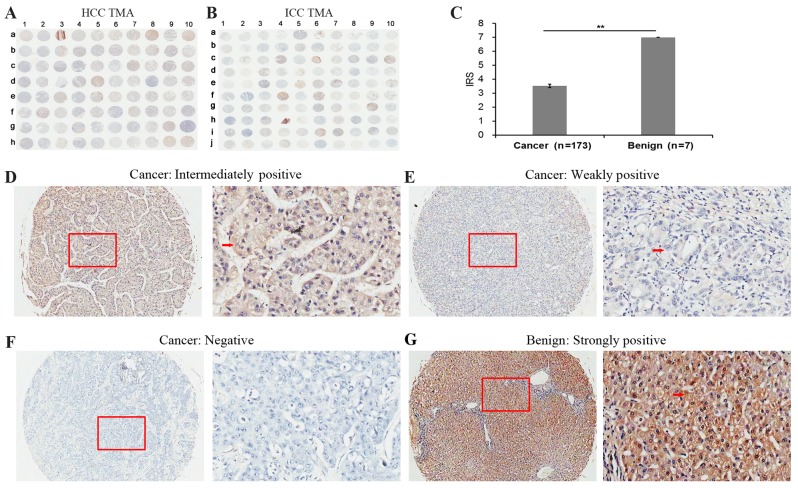
Figure 1.
Immunohistochemical staining for SOX7 in liver cancer and adjacent non-cancerous liver tissues in a TMA. Immunohistochemistry staining for SOX7 in the (A) HCC and (B) ICC TMA cohorts. (C) IRS of SOX7 in liver cancer were lower than those observed in the adjacent normal liver tissues. IRS: **P<0.001, liver cancer (3.53±1.57) vs. benign (7.00±0.00). Data are presented as the mean ± standard deviation. Immunohistochemistry staining indicated that SOX7 immunostaining primarily occurred in the cytoplasm and cellular membrane of liver cancer tissues cells. The intensities of SOX7 immunostaining were (D) intermediate, (E) weak and (F) negative, no strong staining was observed in cancer samples. Left image, magnification ×100; right image, magnification ×400; the red squares indicate the area shown at higher magnification. (G) Strongly positive immunohistochemistry staining in the adjacent non-cancerous liver tissue cells. Red arrows indicate positively-stained cells. SOX7, sex determining region Y-box 7; HCC, hepatocellular carcinoma; TMA, tissue microarrays; ICC, intrahepatic cholangiocarcinoma; IRS, immunoreactivity scores.

