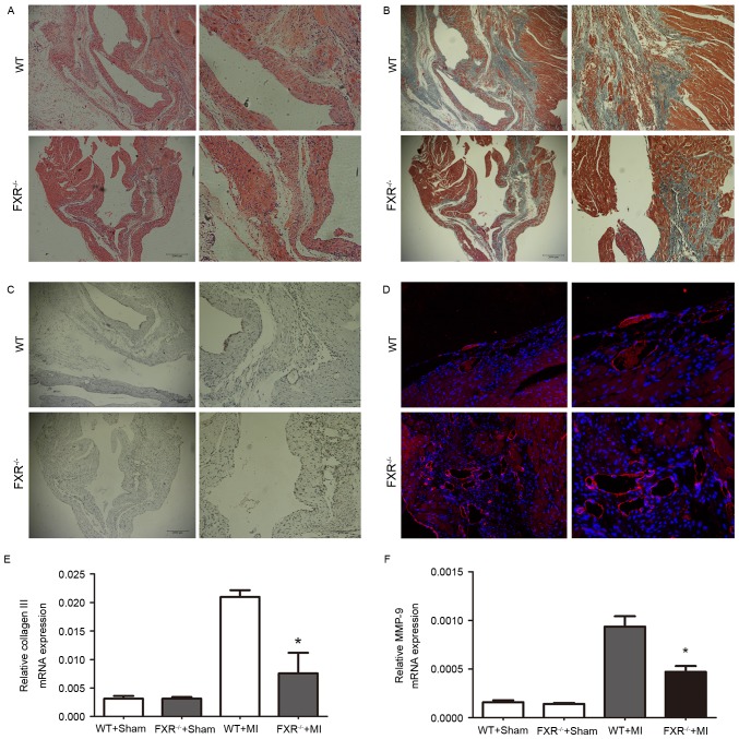
Figure 3.
Decreased interstitial fibrosis and increased microvesicular density in the peri-infarct border zone of FXR−/− mice 4 weeks following MI. (A) Representative photomicrographs (×100 and ×200 magnification) of serial sections stained with hematoxylin and eosin for the morphological identification of the border zone and infarct areas. (B) Representative photomicrographs (×100 and ×200 magnification) of Massons trichrome-stained sections, captured on week 4 following MI, reveal interstitial fibrosis in the border zone in WT and FXR−/− mice. (C) Representative photomicrographs (×100 and ×200 magnification) of sections from the infarcted LV regions of WT and FXR−/− mice stained for α-smooth muscle actin. (D) Representative photomicrographs (×200 and ×400 magnification) of cardiac sections from WT and FXR−/− mice stained with anti-cluster of differentiation 31 on day 28 following MI. mRNA expression levels of (E) collagen III and (F) MMP-9 in LV tissue samples were assessed using reverse transcription-quantitative polymerase chain reaction and normalized to GAPDH expression on day 28 following MI. Data are expressed as the mean ± standard error of the mean (n=6 mice/group). *P<0.05 vs. the WT + MI group. FXR, farnesoid X receptor; MI, myocardial infarction; WT, wild type; MMP, matrix metalloproteinase; LV, left ventricular.

