
Figure 3.
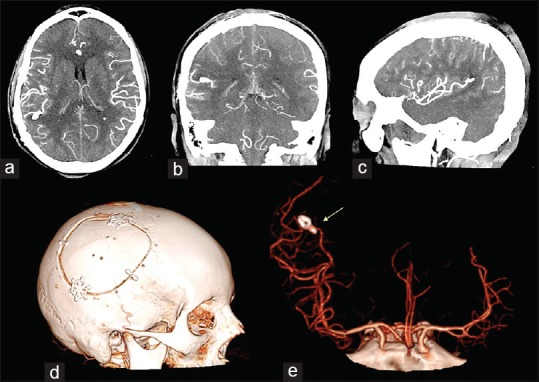
Postoperative intracranial angio-CT scan in axial (a), cronal (b) and sagittal (c) projections showed the results of the aneurysm clipping and the absence of the parietal intraparenchymal haemorrhage. 3D cranial CT rendering of the skull (d) documented the features of the bone flap. 3D angio-CT rendering in antero-posterior ad lateral projection (e) showed the position of the clip and the complete exclusion of the aneurysm (yellow arrow)