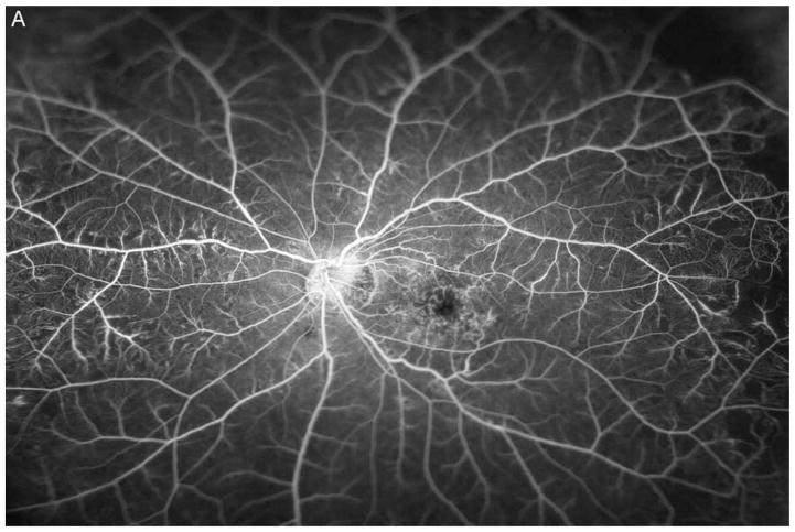
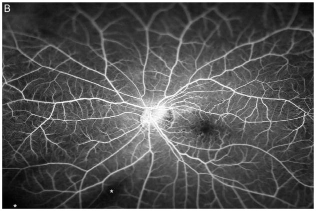
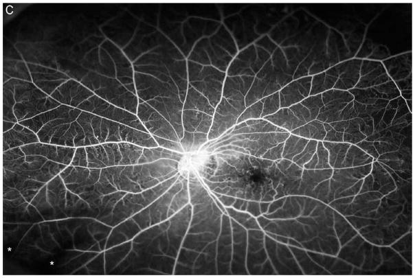
Figure 4. Reduction in area of nonperfusion during monthly ranibizumab injections and partial recurrence of nonperfusion during pro re nata injections in a patient with central retinal vein occlusion.
(A) Image from early recirculation phase of an ultra-wide field fluorescein angiogram (FA) in patient with central retinal vein occlusion shows large patches of RNP in the temporal periphery and small patches in the mid-periphery nasally. (B) Image from early recirculation phase of FA at month 6 after monthly injections of 0.5mg RBZ shows improvement in RNP with many of the areas of RNP in the temporal periphery smaller than baseline. However, there are still small patches of RNP nasally. (C) Image from early recirculation phase of month 12 FA after 6 months of prn RBZ injections, shows worsening of RNP temporally and nasally *artifacts secondary to eye lashes/eyelids.


