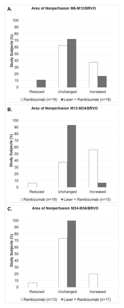
Figure 5. Change in area of retinal nonperfusion in patients with branch retinal vein occlusion in 3 sequential time periods after randomization to pro re nata ranibizumab or ranibizumab+laser.
At month 6, patients with branch retinal vein occlusion (BRVO) were re-randomized to pro re nata (prn) ranibizumab or prn ranibizumab+laser. Ultra-wide field fluorescein angiograms (FA) were used to grade the change in area of retinal nonperfusion as reduced, unchanged, or increased between M6 and month 12 (M6–M12, A), between M12 and month 24 (M12–M24, B), and between M24 and month 36 (M24–M36, C). The distribution among the 3 grades (reduced, unchanged, or increased) was compared by Fisher’s exact test and there was no significant difference in ranibizumab versus laser+ranibizumab groups in the M6–M12 period (A), but significant differences were present in the M12–M24 period (B, p=0.003) and the M24–M36 period (C, p=0.04).

