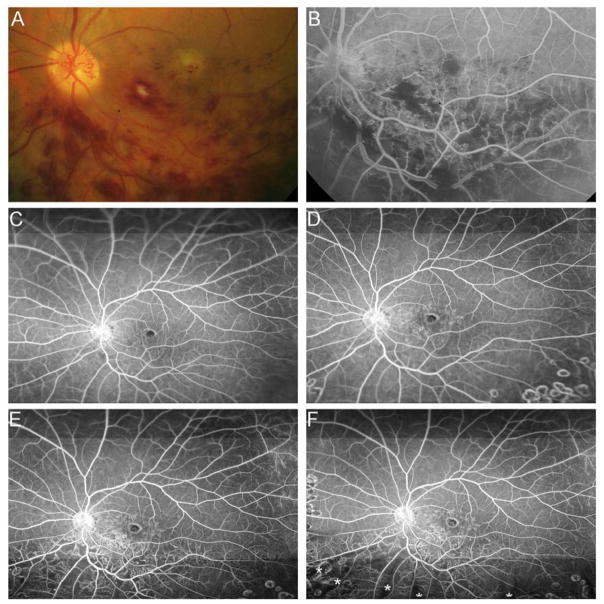
Figure 7. Reperfusion of nonperfused retina in a patient with branch retinal vein occlusion during monthly injections of ranbizimab and failure to prevent recurrent nonperfusion by scatter photocoagulation and pro re nata ranibizumab.
(A) Baseline 30° fundus photograph of patient with inferior branch retinal vein occlusion (BRVO) showing some hemorrhages in the inferior macula. (B) Early recirculation phase of a 30° fluorescein angiogram shows blocked fluorescence corresponding to hemorrhages and areas of retinal nonperfusion (RNP) inferior to the optic nerve. (C) Early recirculation phase of an ultra-wide field (FA) at month 6 after monthly injections of 2.0mg ranibizumab for 6 months shows reperfusion of some previously nonperfused areas. (D) Early recirculation phase of FA at month 12, after 6 months of prn ranibizumab+laser, show no change in RNP (E). Month 24 FA shows a marked increase in RNP inferior and superior to the inferotemporal arcade vessel (F). Month 36 FA shows persistent RNP that was unchanged from the previous visit. (*) Asterisks indicate artifacts secondary to eye lashes/eyelids.

