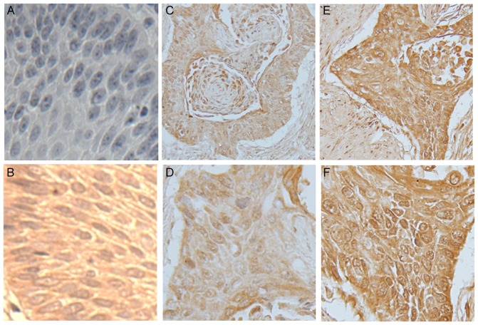
Figure 1.
Immunostaining of PAX9 in normal esophageal mucosa and esophageal squamous cell carcinoma tissue. Representative images of (A) negative control for PAX9 in normal mucosa, (B) positive control for PAX9 in normal mucosa, (C and D) low expression of PAX9 in tumor tissue (low rate of positive cells and low staining intensity, score ≤3) and (E and F) high expression of PAX9 in tumor tissue (high rate of positive cells and high staining intensity, score ≥4). The stained sections were observed and images were captured at low magnification (×100) in C and E, and at high magnification (×200) in A, B, D and F. PAX9, paired box 9.

