
Fig. 1.
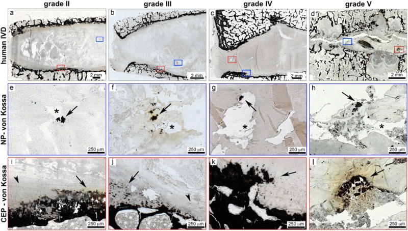
Ectopic calcifications were located at ossification sites and close to fissures in human IVDs of various degeneration stages. Representative overview images of human IVDs of varying degenerative levels including (a) grade II, (b) grade III, (c) grade IV, and (d) grade V. The von Kossa staining identifies ossifications as dark black/brown staining and boxes mark region of interest in NP (blue box) and CEP (red box) regions. In NP regions (e-h), calcified deposits were located close to fissures. Arrows denote calcified deposits and asterisks denote fissures in NP tissues. In CEP regions (i-l), irregular ossified structures were also identified in all degenerative grades. Arrows denote calcified deposits and arrow heads denote intact CEP.