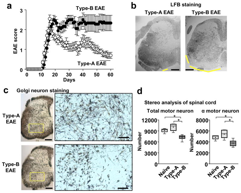
Figure 6. Persistent disease severity and spinal transected neurites in Type-B EAE mice.
(a) EAE scores in WT mice after Type-A and Type-B EAE induction (n=9). (b) Representative LFB-stained images of spinal cord sections. Yellow lines indicate regions of demyelination. (c) Representative silver staining images to show neurons in spinal cord sections. Areas within the yellow rectangles were enlarged. Scale bars in (b) and (c, original size picture), 200 μm, and in (c, magnified pictures), 50 μm. (d) Motor neuron stereology. Total Nissl+ and α-motor neuron in lumber region of the spinal ventral horn were evaluated. *; p<0.05. Total motor neuron number: P=0.0001, F20.61. α motor neuron number: P=0.0032, F=11.65, by Bonferroni’s Method (n=6). All the CNS samples were harvested on 70-dpi. All the experimental data and images are representatives from at least 2 similar experiments for each.

