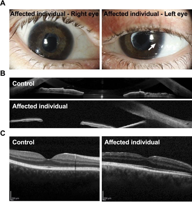
Figure 6.

(A) Slit-lamp images of the eyes of an affected human individual with SHORT syndrome revealing embryotoxon posterior (arrow) and iris atrophy. In addition, two iridotomies after previous trabeculectomies can be seen in the upper part of the iris in the right eye, and anterior capsule opacification after cataract surgery in both eyes. (B) A representative OCT image showing the iris of an unaffected individual (top) and an affected individual (the son) with SHORT syndrome (bottom), showing thinner irides in the affected compared to the unaffected individual. (C) A representative OCT image showing the retina of an unaffected individual (left) and the retina of the same affected individual as above (the son) with SHORT syndrome, showing thinning of the nerve fiber layer. The pigment epithelium appeared poorly pigmented.
