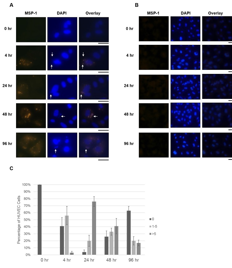
Figure 3.
Detection of P. falciparum associated with HUVECs by MSP-1 staining. P. falciparum iRBCs (A) or uninfected RBCs (B) were added to HUVEC cell cultures for the indicated times, as described in Materials and Methods. A. Following incubation with HUVECs, P. falciparum-derived particles were detected by immunofluorescence assay with α-MSP1 primary antibody (first column). DAPI staining shows nucleic acid of HUVECs and parasite-like bodies (indicated by arrows, second column). Overlays of MSP1 and DAPI staining shows overlap of MSP1 and DAPI fluorescence (arrows). B. HUVECs incubated with uninfected RBCs showed no spots of bright fluorescence from MSP1 staining (first column). DAPI staining shows HUVEC nucleic acid, but no smaller spots corresponding to parasite nucleic acid were observed (second column). The black bars below the micrographs indicated 25 μm. C. The percentage of HUVEC cells containing 0, 1 to 5, or > 5 P. falciparum-derived particles was calculated for each time point.

