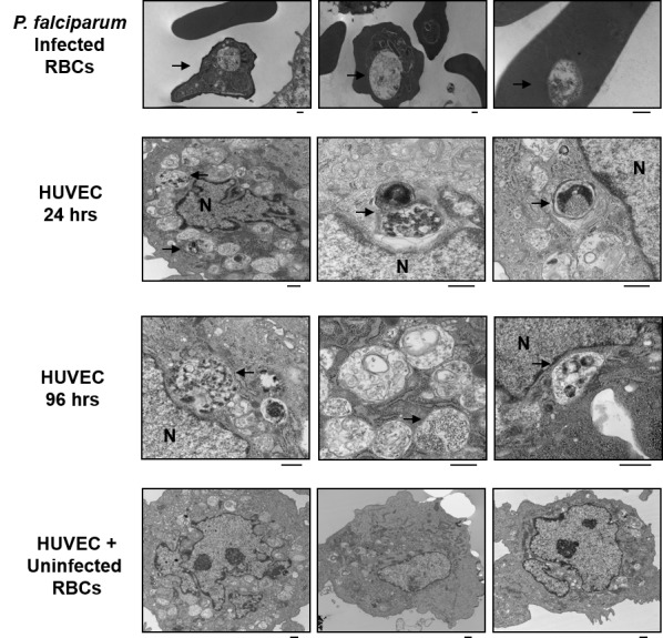
Figure 5.

Visualization of HUVECs after incubation with iRBCs by electron microscopy. (Top row) P. falciparum cultures were grown in RBCs and fixed for electron microscopy. (Second row) HUVEC cultures were incubated with RBCs infected with GFP-expressing P. falciparum for 24 hours. (Third row) For 96 hr cultures, RBCs were removed after 24 hours, fresh medium was added, and cultures were incubated an additional 72 hours. (Bottom row) Aberrant structures seen only in HUVECs incubated with iRBCs are indicated by arrows. As a control, HUVECs were incubated with uninfected RBCs for 24 hours. No such aberrant structures were seen in these cells. HUVEC nucleus is indicated (N). Black scale bar represents 500 nm.
