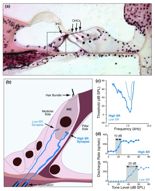
Figure 2. High- vs. low-SR auditory nerve fibers and their synaptic localization on the inner hair cell.
(a) Light micrograph of the organ of Corti, as it appears in conventional histological material, stained with hematoxylin and eosin. Peripheral terminals of auditory nerve fibers (ANFs) in the inner hair cell (IHC) area (box) are not resolvable. (b) Schematic of type I peripheral terminals showing that fibers with high versus low spontaneous discharge rates (SRs) make synaptic contacts on opposite sides of the IHC. (c) High-SR fibers have lower thresholds than do low-SR fibers, as shown by these two tuning curves. (d) High-SR fibers have smaller dynamic ranges (grey box) than do low-SR fibers when stimulated with tone bursts at the characteristic frequency. dB SPL, decibels sound pressure level; OHC, outer hair cell.

