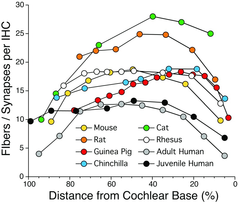
Figure 4. Normal density of auditory nerve fibers along the cochlear spiral.
Data from the mouse, rat, guinea pig, chinchilla, rhesus monkey, and adult human are from the Liberman lab and are based on confocal analysis of immunostained synapses from cochlear epithelial whole mounts such as in Figure 5. Cat data are from a serial-section ultrastructural study 77. Data from juvenile human are based on light-microscopic counts of peripheral axons from a 7-year-old 42. Deviation between the two sets of human data at low frequencies may arise because ANFs in apical cochlear regions often form two synapses each 11.

