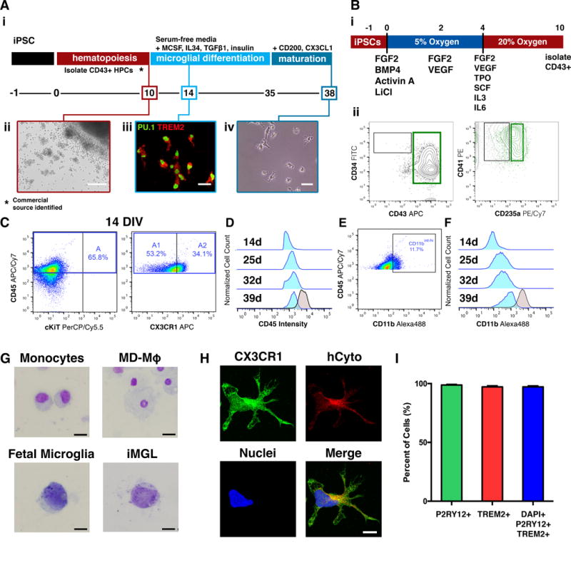
Figure 1. Differentiation of human iPSC derived microglia like cells (iMGLs).

(A) Schematic of fully-defined iMGL differentiation protocol. (i) Human iPSCs are differentiated to CD43+ iHPCs for 10 days and then cultured in serum-free microglia differentiation media containing human recombinant MCSF, IL-34, and TGFβ-1. Differentiation is carried out for an additional 25 days after which iMGLs are exposed to human recombinant CD200 and CX3CL1 for 3 days. (ii) Representative image of iHPCs in cell culture at day 10. Scale bar = 100 μm. (iii) By day 14, iMGLs express PU.1 (green) and TREM2 (red). Scale bar = 50 μm. (iv) Representative phase contrast image of iMGL at day 38. (B) Schematic of differentiation of iPSCs to iHPCs. (i) Single-cell iPSCs are differentiated in a chemically defined media supplemented with hematopoietic differentiation factors and using 5% O2 (4 days) and 20% O2 (6 days). (ii) After 10 days, CD43+ iHPCs are CD235a+/CD41a+(C) iMGLs develop from CD45+/CX3CR1− (A1) and CD45+/CX3CR1+ (A2) progenitors. (D) CD45 fluorescence intensity shows that iMGLs (blue) maintain their CD45lo-int profile when compared to monocyte-derived macrophages (MD-Mφ). (E) iMGL progenitors are CD11blo and increase their CD11b expression as they mature. At 14 DIV, a small population (~11%) cells with CD11bint-hi are detected. (F) CD11b fluorescence intensity demonstrates that CD11b expression increases as iMGLs age, resembling murine microglial progenitors identified by Kierdorf, et al 2013. (G) Mary-Grunwald Giemsa stain of monocytes, MD-Mφ, fetal microglia, and iMGLs. Both fetal microglia and iMGL exhibit a high nucleus to cytoplasm morphology compared to monocytes and MD-Mφ. Scale bars = 16 μm. (H) iMGLs also exhibit extended processes and express CX3CR1 (green) together with the human cytoplasmic marker (hCyto, SC121; red). (I) Differentiation yields >97.2% purity as assessed by co-localization of microglial-enriched protein P2RY12 (green), microglial-enriched TREM2 (red) and nuclei (blue (n=5 representative lines). See also Figures S1 and S2.
