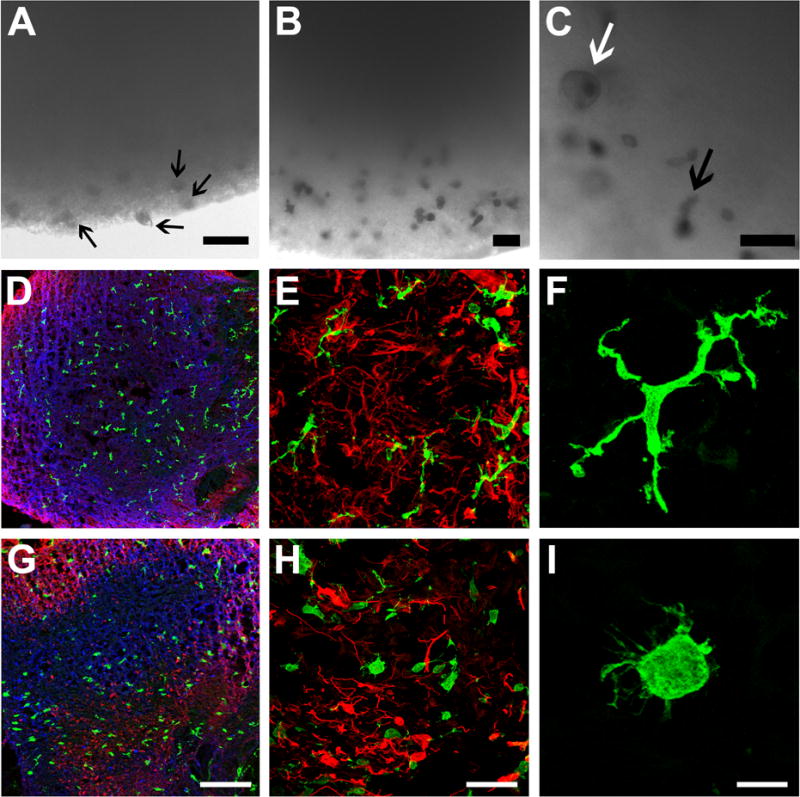
Figure 6. iMGLs respond to the neuronal environment in 3D brain organoid co-cultures (BORGS).

iMGLs (5 × 10⁵ cells) were added to media containing a single BORG for 7 days. (A) Representative bright-field image of iMGLs detected in and near BORG after 3 days. iMGLs were found in and attached to the BORG-media interface (arrows), but not free floating in the media, suggesting complete chemotaxis of iMGLs. (B) Representative image of iMGLs in outer and inner radius of BORG. (C) Embedded iMGLs exhibit macrophage-like morphology (white arrow) and extend processes (arrow) signifying ECM remodeling and surveillance respectively. Simultaneous assessment of embedded iMGL morphology in uninjured (D–F) and injured (G–I) BORGs. (D) Immunohistochemical analysis of BORGs reveals iMGLs begin tiling evenly throughout the BORG and project ramified processes for surveillance of the environment. BORGs are representative of developing brains in vitro and contain neurons (β3-tubulin, blue) and astrocytes (GFAP, red), which self-organize into a cortical-like distribution, but lack microglia. iMGLs (IBA1, green). (E–F) Representative immunofluorescent images of iMGLs with extended processes within the 3D CNS environment at higher magnification. (G–I) Representative images of iMGL morphology observed in injured BORG. (H–I) Round-bodied iMGLs reminiscent of amoeboid microglia are distributed in injured BORGs and closely resemble activated microglia, demonstrating that iMGLs respond appropriately to neuronal injury. Scale Bar (A–C) =50 μm in A-C, (D,G)= 200 μm (E–H)= 80 μm and (F,I) =15 μm.
