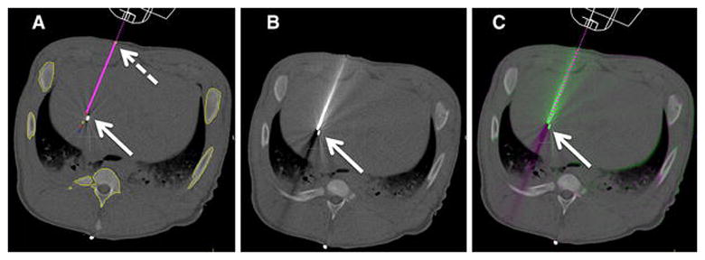
Figure 2. Procedure Workflow.

A: Planning image, magenta line shows planned needle trajectory from skin entry (dashed arrow) to target seed (arrow).
B: Post needle placement CT image.
C: Image overlay comparing planned trajectory (dashed line) and actual needle position.
