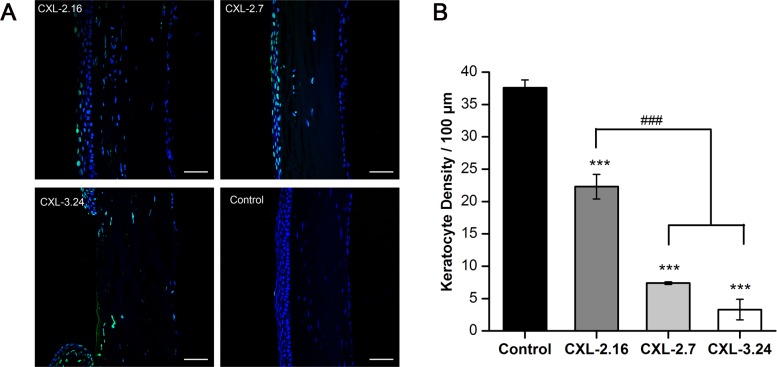
Fig 5. CXL-induced apoptosis and keratocyte density.
(A) Apoptosis of the rat keratocytes (green, positive TUNEL staining) are present in the CXL-treated areas and control cornea on day 3. The control cornea had a normal DAPI staining pattern. In the cross-linked corneas, some keratocyte were apoptotic. Apparent endothelial layer damage was detected in the CXL-3.24 group. (B) Significant difference was evident between keratocyte density in CXL-treated and control corneas on day 3. The keratocyte counts significantly decreased in the CXL-2.7 and 3.24 groups compared with the CXL-2.16 group (### p < 0.01 versus CXL-treated groups, *** p < 0.001 versus control group, respectively; Data are presented as Mean ± SEM, n = 3. scale bars = 50 μm).

