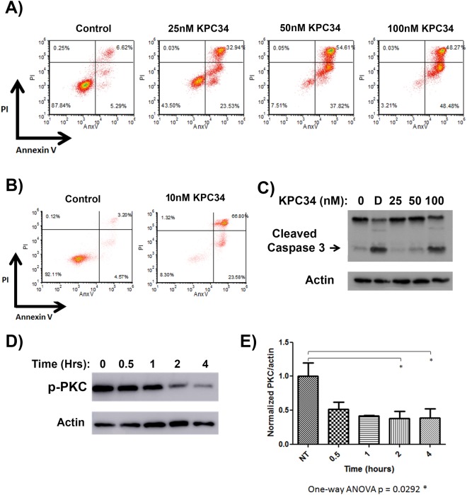
Fig 1. KPC34 induces apoptosis in a dose-dependent fashion and inhibits PKC phosphorylation.
- A) Flow cytometry of annexin V assay. B6-ALL cells were treated with the indicated amount of KPC34 for 48 hours and then labeled with annexin V and propidium iodide (PI).
- B) Flow cytometry of annexin V assay. SUPB15 cells were treated with the indicated amount of KPC34 for 48 hours and then labeled with annexin V and propidium iodide (PI).
- C) Western blot analyses of apoptotic induction. B6-ALL cells were treated with the indicated amount of KPC34 for 48 hours or for four hours with 500 ng/ml doxorubicin (D) as a positive control and blotted for caspase 3. Arrow denotes cleaved caspase 3. Actin was used as a loading control.
- D) Western blots of phosphorylated PKCα and βII. B6-ALL cells were treated with 200 nM KPC34 for the indicated time and were blotted with an antibody that recognizes both phosphorylated PKCα and PKCβII (p-PKC). Actin was used a loading control.
- E) Quantified p-PKC expression of B6-ALL cells treated with vehicle or KPC34 200 nM. Band intensities from three independent experiments were calculated using ImageJ software and p-PKC values were normalized to actin for each replicate. Errors bars represent the SEM.

