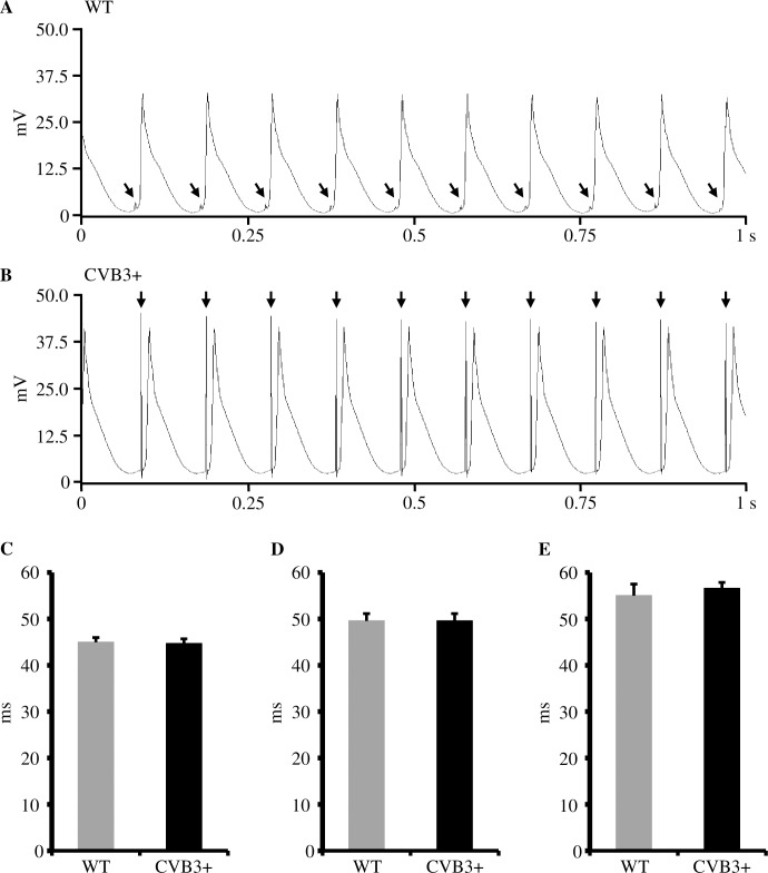
Fig 3. Monophasic action potentials and action potential duration (APD90) in WT and CVB3+ whole hearts.
(A) Representative recording of monophasic action potentials in a Langendorff-perfused WT whole heart at pacing with S1 = 100 ms (ms = milliseconds). mV = millivolt, s = seconds. ↓ = electrical stimulus artefact. (B) Representative recording of monophasic action potentials in a Langendorff-perfused CVB3+ whole heart at pacing with S1 = 100 ms. (C) APD90 in Langendorff-perfused WT and CVB3+ hearts at pacing with S1 = 100 ms. WT n = 7; CVB3+ n = 7. n = number of Langendorff-perfused hearts. (D) APD90 in Langendorff-perfused WT and CVB3+ hearts at pacing with S1 = 120 ms. WT n = 7; CVB3+ n = 7. (E) APD90 in Langendorff-perfused WT and CVB3+ hearts at pacing with S1 = 150 ms. WT n = 6; CVB3+ n = 7. Within each genotype, we found a heart rate dependent shortening of ADP90 with increasing heart rate. (WT ADP90 at: S1 = 100 ms vs. S1 = 150 ms, p = 0.014; CVB3+ ADP90 at: S1 = 100 ms vs. S1 = 150 ms, p = 0.001).

