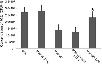
Fig. 5.

Growth of M.tb in THP-1-derived macrophages. Results are expressed as CFU/ml. (1) M.tb, M.tb-infected macrophages were maintained in RPMI1640 culture medium for 6 days; (2) M.tb+SD(CTL), M.tb-infected macrophages were maintained in RPMI1640 culture medium containing DMSO (the solution medium of SD) for 6 days. DMSO was added to the culture medium at the same volume as SD; (3) M.tb+SD, M.tb-infected macrophages were maintained in RPMI1640 culture medium containing 6 μM SD for 6 days; (4) M.tb+SD+DOR(CTL), M.tb-infected macrophages were maintained in RPMI1640 culture medium containing 6 μM SD and the solution medium of DOR(DMSO) for 6 days. The solute medium of DOR was added to the culture medium based on the volume of DOR; (5) M.tb+SD+DOR, M.tb-infected macrophages were maintained in RPMI1640 culture medium containing 6 μM SD and 1 μM DOR for 6 days. Data are shown as the mean ± SD of 4 independent experiments carried out from 6 parallel infections. ★P < 0.05 versus M.tb + SD group
