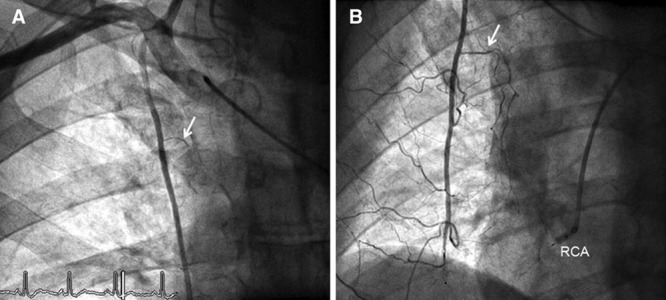
Figure 1.

A, Anteroposterior angiogram of the truncus brachiocephalicus (site of the catheter tip) depicting the right internal mammary artery (RIMA) with its pericardiophrenic branch (arrow) taken at the baseline examination. B, RIMA angiography of the same patient as in A taken during follow-up examination. Simultaneous occlusion of the distal RIMA (by vascular occlusion device) and the ostial right coronary artery (RCA, by angioplasty balloon). The pericardiophrenic branch (arrow) and the other RIMA branches are larger than those at baseline (A).
