
Figure 8. The expression of BDNF protein in the hippocampus of CUMS rats upon the activities of COX2.
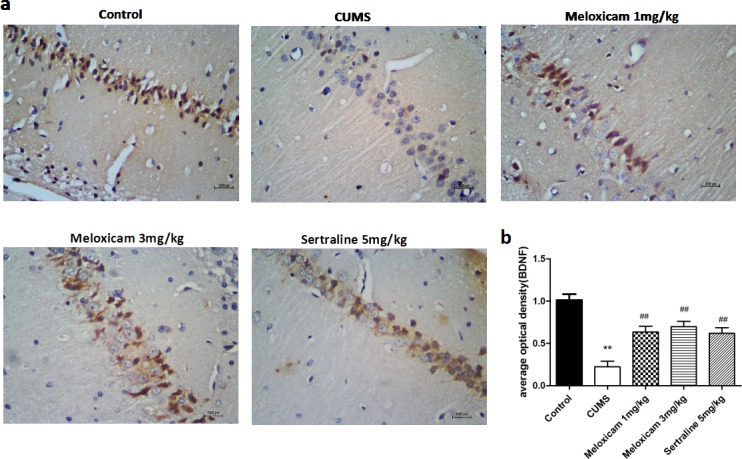
a. Representative immunohistochemical staining for BDNF positive areas in the hippocampal brain sections from CUMS rats after meloxicam (1 and 3mg/kg, i.g.) or sertraline (5mg/kg, i.g.) treatment. b. Densitometric analyses of the immunoreactivity to the BDNF antibodies from the previous panel Figure 8 (a). Positive cells are represented as browns spots. Scale bar = 100 um. The level of staining density was quantified by Image-ProPlus 6.0 and presented as the mean ± SD using a one-way ANOVA with a Bonferroni correction. **P < 0.01 vs control group. # and ##P < 0.05 and < 0.01 vs the CUMS group. n = 10.