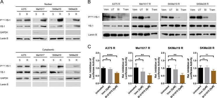
Figure 5. YB-1 activity is increased in vemurafenib resistant melanoma cells as a consequence of elevated MAPK/RSK signalling.
(A) Immunoblot analysis of total and S102-phosphorylated YB-1 in cytoplasmic and nuclear enriched fractions of sensitive and vemurafenib resistant melanoma cells. GAPDH and Lamin B served as the respective subcellular markers. One representative experiment is shown (n = 2 or n = 3). (B) Western Blot analysis of total and PS102-YB-1 in nuclear enriched fractions of vemurafenib resistant cells treated with vemurafenib (2 μM), trametinib (50 nM), BI-D1870 (5 μM) or left untreated (UT) for 24 h. Lamin B served as loading control. (C) (Y-box)4-luc luciferase reporter assay reflecting YB-1 transcriptional activity in vemurafenib resistant melanoma cells after a 24 h-treatment with MAPK/RSK pathway inhibitors. Firefly luciferase activity was normalized to the protein content and its relative inhibition in response to treatment presented in the graph (n = 5; mean ± SD). Significance was determined with 1-way ANOVA and subsequent Tukey's multiple comparison test.

