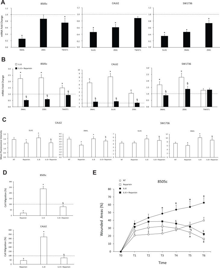
Figure 3. Reparixin inhibits the epithelial-to-mesenchymal transition (EMT) in TC cells.
(A–B) Effect of Reparixin (30 μM) on mRNA levels of the basal and IL-8 induced-EMT markers (SLUG, SNAIL, ZEB1, TWIST1) was evaluated by real time PCR in 8505c, CAL62 and SW1736 cells. Normalization to β-actin mRNA levels was used. For each target (x-axis), the expression levels were calculated relative to the expression levels in untreated cells, arbitrarily considered equal to 1 (dotted line). Experiments were performed in triplicate. *p < 0.05 compared to untreated cells (dotted line); §p < 0.05 compared to IL-8 treated cells. (C) Cytofluorimetric evaluation of the indicated EMT markers levels in Reparixin (30 μM)-treated CAL62 and SW1736 cells, in the presence or absence of IL-8 (100 ng/ml). *p < 0.05 compared to untreated cells (NT); §p < 0.05 compared to IL-8 treated cells. (D) Migration assay on Reparixin (30 μM)-treated 8505c and CAL62 cells, in presence or absence of IL-8 (100 ng/ml). The results were expressed as percentage of migrated cells compared to control (dotted line). *p < 0.05 compared to untreated cells (dotted line); §p < 0.05 compared to IL-8 treated cells. (E) Wound-healing assays of 8505c cells treated with Reparixin (30 μM) in presence or absence of IL-8 (100 ng/ml) assessed in a time course experiment (0–18 h, every 3 h). Wound closure was measured by counting migrated cells in the wound area by high content analysis (Operetta, PerkinElmer). Experiments were performed in triplicate. *p < 0.05 compared to untreated cells (NT); §p < 0.05 compared to IL-8 treated cells.

