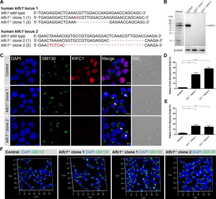
Figure 4. Kifc1 knockout also results in the disorganization and dispersal of the Golgi apparatus.
(A) Representative sequences of the human kifc1 locus targeted by Cas9n and the selected sequences showing representative indels. Generation of indels in the human kifc1 locus in HEK293T cells by CRISPR-Cas9 system. Two different kifc1 knockout cell lines were selected as candidates in this study (designated as kifc1−/− clone 1 and clone 2). (B) Western blot analysis of KIFC1 expression in control, kifc1−/− clone 1 and clone 2 HEK293T cells. β-Actin was used as the loading control. (C) Representative confocal images of the Golgi marker GM130 and KIFC1 in control and kifc1 knockout HEK293T cells (kifc1−/− clone 1 and clone 2). DAPI, blue; GM130, green; KIFC1, red. DIC, differential interference contrast. Scale bars: 5 μm. (D) Quantification of the ratios of disorganized Golgi apparatus in control and kifc1 knockout HEK293T cells (n = 3 cultures for each phenotype). Data are presented as means ± SEM. ns, not significant; *P < 0.05; **P < 0.01; ***P < 0.001 (Student's t-test). (E) Quantification of the ratios of the dispersal Golgi in control and kifc1 knockout HEK293T cells (n = 3 cultures for each phenotype). Data are presented as means ± SEM. ns, not significant; *P < 0.05; **P < 0.01; ***P < 0.001 (Student's t-test). (F) Representative three dimensional images of the Golgi apparatus (DAPI, blue; GM130, green) in the control, kifc1−/− clone 1 and clone 2 HEK293T cells.

