
Figure 7.
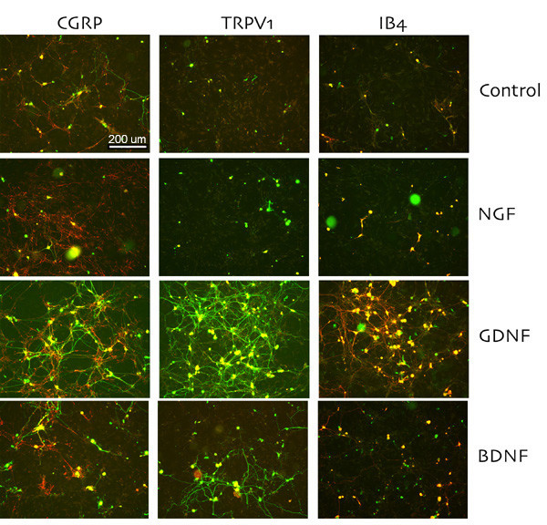
Representative 20X photomicropgraphs of colocalization of sensory neurons markers with NF-H in growth factor-treated TG neurons. Immunoreactivity for CGRP and TRPV1 and staining for IB4-binding sites (red) was assessed following immunocytochemistry for NF-H (green) to assess the proportion of neurons expressing these population markers. Representative 20X photomicrographs of each growth factor at 100 ng/ml for CGRP (left), TRPV1 (middle) and IB4 (right) are shown as well as control (no growth factor-treated) TG cultures (top panels). All cell bodies containing both NF-H-immunoreactivity and CGRP- or TRPV1-immunoreactivity or IB4-binding appear yellow from the overlay of the red with green. In no cases were neurons observed that contained CGRP- or TRPV1-immunoreactivity or IB4-binding without the co-presence of NF-H-immunoreactivity.