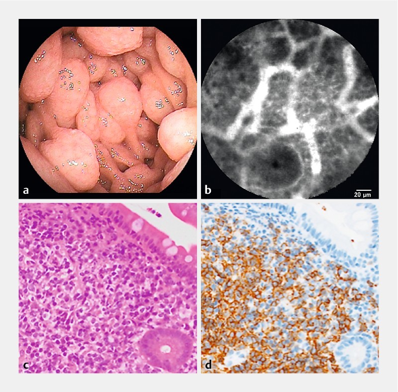
Fig. 2.

Mantle cell lymphoma (Patient #1). a Enteroscopic view in the jejunum. b A probe-based confocal laser endomicroscopic image shows the homogenous cells packed with polygonal capillary vessels in a “soccer ball-like pattern. c H&E staining in a biopsy specimen with a magnification of × 200. d Immunohistochemical staining with CD20 antibodies in a biopsy specimen with a magnification of × 200.
