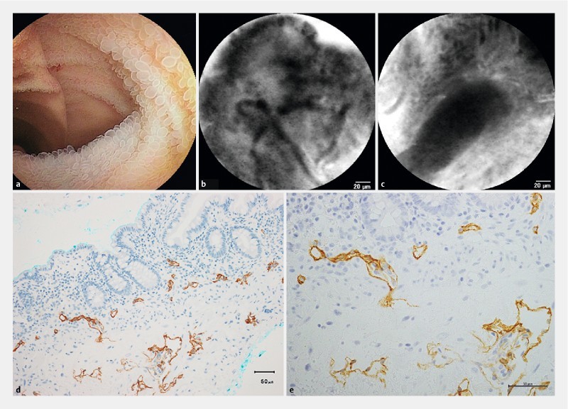
Fig. 7.

Intestinal lymphangiectasia, non-white type with protein-losing enteropathy (Patient #30). a Enteroscopic view with small round villi in the ileum. b A probe-based confocal laser endomicroscopic image on the surface of the villus with unenhanced lymphatic vessels. c A probe-based confocal laser endomicroscopic image deep in the mucosal layer with an unenhanced lymphatic duct. d Immunohistochemical staining with D2-40 antibodies in a biopsy specimen. Magnification × 200. e Immunohistochemical staining with D2-40 antibodies in a biopsy specimen. Magnification × 400.
