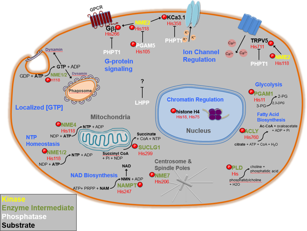
Figure 2. Summary of pHis Cellular Functions.
An illustration of the pHis related proteins discussed in this review and their various functions, enzymatic reactions and subcellular localizations. NME1/2 protein histidine kinase functions are in yellow, pHis enzyme intermediates are in green, phosphohistidine phosphatases are in white and pHis substrates are in bold. Beneath each protein’s gene name is the specific amino acid position number of the pHis residue in red. Cellular functions of specific pHis proteins are in blue. The subcellular localization of pHis related proteins and functions are in grey. Curved arrows represent reactions catalyzed by enzymes that utilize pHis intermediates. For LHPP, phospholysine and 3-phsphohistidine are substrates in vitro, however no known substrates have yet been identified in vivo.

