Figure 3.

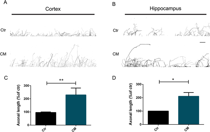
Axonal-specific stimulation with CM induces axonal growth of CNS neurons. (A,B) Effect of local application of CM on axonal outgrowth. Cortical (A) and hippocampal (B) axons present in the axonal compartment were stimulated with CM for 24 h at DIV5-6. Axonal outgrowth was evaluated by immunocytochemistry using anti-tubulin βIII. The area selected comprises 3 mm of chamber length which comprises the area between the first and the last microgrooves. Contiguous images were taken using an AxioObserver Z1 fluorescent microscope with a PlanApochromat 20× objective and assembled into a single image using the ZEN 2011 software. (C,D) Quantification of axonal length. Results show that in the presence of CM the axonal network significantly increases comparatively to control, demonstrating that specific local application of CM to cortical and hippocampal axons promotes axonal outgrowth. Axonal network was measured using Neurolucida software. Bars represent the mean ± SEM of 3 independent experiments. *Represents p = 0.0286 by Mann Whitney unpaired t-test when compared to Ctr. (C) **Represents p = 0.0079 by Mann Whitney unpaired t-test when compared to Ctr. The scale bar is 250 µm.
