Figure 6.

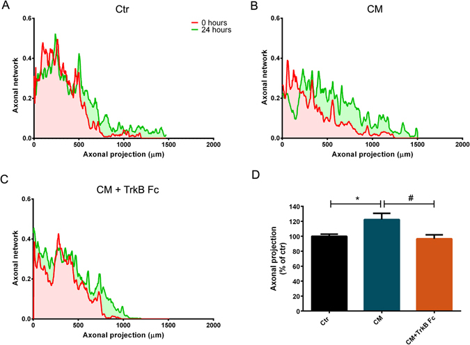
BDNF is the molecule responsible for CM-induced axonal elongation in distal cortical axons. (A–C) Quantification of axonal outgrowth with the MATLAB script AxoFluidic. Profile along the xx axis of the microfluidic device showing the area occupied by the axons at 0 hours (clear red area) and at 24 hours (clear green area). Red line represents axonal projection at 0 hours and the green line represents axonal projection at 24 hours. (D) Quantification of axonal projections outgrowth between 0 and 24 hours. Distal axons treated with CM for 24 hours present more pronounced elongation than control or CM-lacking BDNF axons. Bars represent the mean ± SEM of 6 microfluidic chambers randomly selected of 3 independent experiments. *Represents p = 0.0346 by one-way ANOVA analysis of variance using Tukey post test when Ctr is compared to CM; #represents p = 0.0145 by one-way ANOVA analysis of variance using Tukey post test when CM is compared to CM + TrkB Fc.
