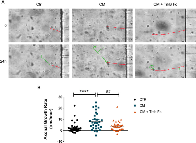
Figure 7.

BDNF works as a localized signal in CM-induced axonal outgrowth. (A) Depletion of BDNF from CM in distal cortical axons abolished CM-induced axonal outgrowth. The axonal outgrowth of individual axons was assessed by live-cell imaging at the moment of CM addition to the axonal compartment of microfluidic chambers and 24 h later. The results show that BDNF depletion results in the abolishment of CM-induced axonal outgrowth in isolated cortical axons, in agreement with the results observed in Fig. 6, demonstrating that BDNF is a key component of CM-induced axonal elongation. Contiguous images were taken using an AxioObserver Z1 fluorescent microscope with a PlanApochromat 20× objective and assembled into a single image using the ZEN 2011 software. (B) Quantification of axonal growth rate. Fresh NBM, CM and CM + Trkb-Fc were added to the axonal compartment at DIV4 and axons allowed to develop for 24 hours. After this period, new images were acquired at the exact same position in the axonal compartment of microfluidic chambers, and the growth rate of individual axons were analyzed with Image J 1.45e software (see Material and methods for details). Results show an increase in axonal growth rate during CM stimulation, however depletion of BDNF blocks the observed axonal growth rate after CM stimulation. Plots represent the mean ± SEM of approximately 30 axons selected of 3 independent experiments. ****Represents p < 0.0001 by one-way ANOVA analysis of variance using Tukey post test when Ctr is compared to CM; ##represents p = 0.0080 by one-way ANOVA analysis of variance using Tukey post test when CM is compared to CM + TrkB Fc. The scale bar is 50 µm.
