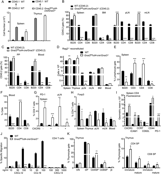
Figure 5.

Reconstitution of WT and Rag2 −/− mice with DKO bone marrow results in different phenotypes. (A) Number of splenocytes and thymocytes recovered from WT reconstituted mice. CD45.1 WT mice were lethally irradiated and reconstituted with bone marrow from CD45.2 WT or CD45.2 DKO mice. Seven weeks later the number of CD45.1 and CD45.2 cells in the spleen and thymus were enumerated. (B,C) The percentage of different cell types in the lymphocyte gate that also expressed CD45.2. Cells were isolated from the spleen, bone marrow (BM), pLN, and mLN (B) and PP and blood (C) from the reconstituted mice. (D) Number of lymphoid cells recovered from Rag2 −/− reconstituted mice. CD45.1 Rag2 −/− mice were lethally irradiated and reconstituted with bone marrow from CD45.2 WT or CD45.2 DKO mice. Seven weeks later the number of CD45.1 and CD45.2 cells in various lymphoid organs were enumerated. (E,F) Lymphocyte subsets in the spleen (E) and blood (F) of Rag2 −/− reconstituted mice. (G) Distribution of PD-1+ CXCR5 positive or negative CD4 T cells in the spleen and pLNs of Rag2 −/− reconstituted mice. (H) Percentage of Foxp3+ CD4 T cells in different lymphoid organs and the blood. (I) Comparison of indicated cell surface markers on CD4 T cells from Rag2 −/− bone marrow reconstituted mice. Data presented as a geometric mean. (J) Chemotaxis assays with increasing concentrations of S1P, CXCL12, and CCL19 as indicated. Shown are the percentage of CD4 T cells from Rag2 −/− mice reconstituted with the indicated bone marrow that specifically migrated. (K) Distribution of thymocyte subsets in Rag2 −/− mice with bone marrow from WT or DKO mice. Results are from 8 mice WT or 8 Rag2 −/− mice reconstituted either with WT or DKO mice. *p < 0.05, **p < 0.005 and ***p < 0.0005 (Student’s t-test).
