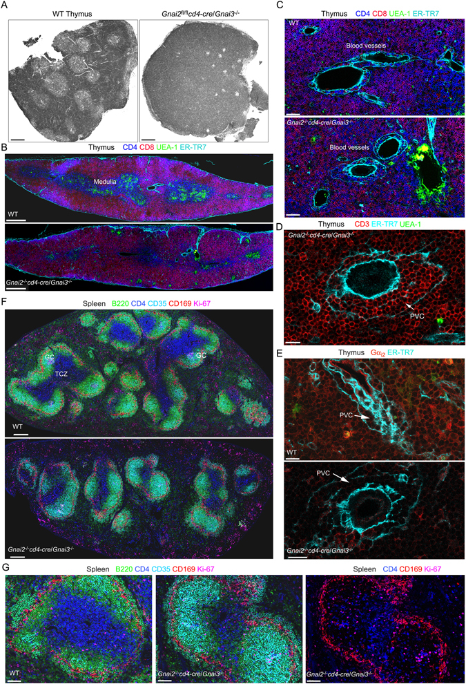
Figure 7.

Consequences of deleting Gnai2 in DP thymocytes of mice that lack Gnai3 on lymphoid organ architecture. (A) Confocal microscopy image collected with a transmission photomultiplier tube of a hematoxylin & eosin stained thymus sections from the indicated mice. Scale bar is 300 μM. (B) Confocal microscopy of a thymus section from the indicated mice immunostained for CD4, CD8, UEA-1, and ER-TR7. Acquired images tiled together to span a single thymus lobe. Scale bar are 400 μM. (C) Electronically zoomed images from part B focused on a likely site of thymocyte egress. Scale bar is 50 μM. (D) Confocal microscopy of a thymus section from a DKO mouse immunostained for CD3, ER-TR7, and UEA-1 focused on a likely egress vessel. The perivascular channel (PVC) is indicated. Scale bar is 15 μM. (E) Confocal microscopy of thymus sections from the indicated mice. The sections were immunostained for Gαi2 and ER-TR7. The PVC is indicated. Scale bars are 15 μM. (F) Tiled confocal images of the spleens of the indicated mice immunostained for B220, CD4, CD35, CD169, and Ki67. The sections are oriented parallel to the short axis of the spleens. Several germinal centers (GC) are evident in the WT spleen. (G) Zoomed image of a splenic follicle of the indicated mice from F. The sections were immunostained for B220, CD4, CD35, CD169, and Ki-67. In the far right image the B220 and CD35 signals have been removed. Scale bars are 60 μM.
