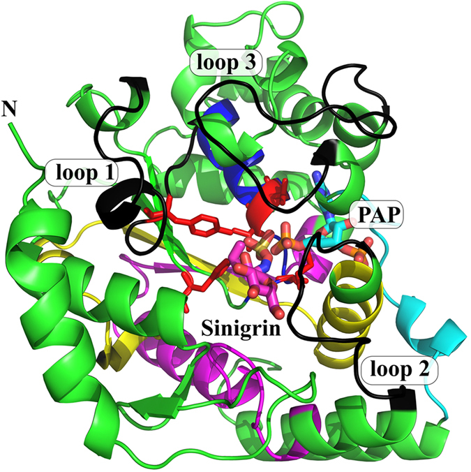
Figure 3.

Overall view of AtSOT18 from two perspectives bound with sinigrin (magenta sticks) and PAP (cyan sticks). Indicated are the four conserved regions (region I: blue; region II: yellow; region III: magenta: region IV: cyan), the three flexible loop regions (black), the catalytic residues (red) and proline 136 (orange). The upper view shows the three flexible loops gating the sinigrin binding site. The view below shows how two of the four typical β-strands are formed by the conserved regions II and III.
