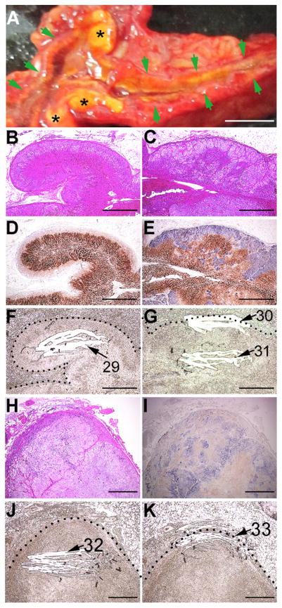
Fig. 2.
Gross, histochemical, and immunohistochemical findings. A: Gross appearance of the left adrenal cut surface. Green arrows and black asterisks (*) indicate a normal adrenal gland portion and hyperplastic/adenomatous portions, respectively. B, C, and H: H&E staining. Enlarged images of the boxes in pages#1 and #4 of Supplementary Fig. 1. D, E, and I: Double immunostaining for CYP11B2 (blue) and CYP11B1 (brown). Enlarged images of the boxes in pages#1 and #4 of Supplementary Fig. 1. F, G, and J/K: Macro-dissected unstained adjacent FFPE sections of panels D, E, and I, respectively. Dotted lines indicate the adrenal capsule. White striations and corresponding numbers indicate scraped (macro-dissected) areas and DNA# in Table 1, respectively. The scale bar in panel A indicates 5 mm. Scale bars in panels B–K indicate 1 mm. (For interpretation of the references to colour in this figure legend, the reader is referred to the web version of this article.)

