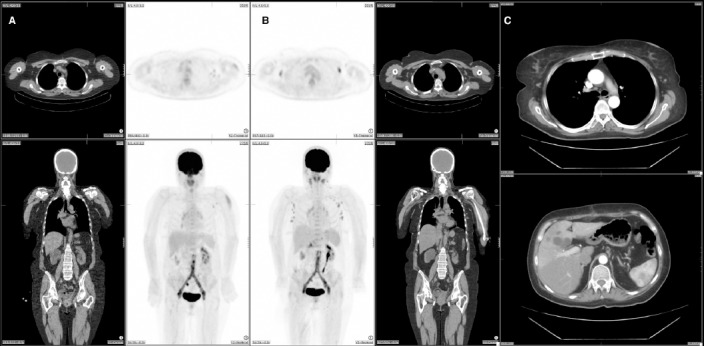
Figure 1.

The first FDG-PET/CT study shows activity in the left deltoid and ipsilateral axillary lymph nodes (A). The second PET/CT study shows less but persistent FDG activity around the aortic graft as well as multiple FDG-avid lymph nodes on both sides of the diaphragm including bilateral cervical, bilateral axillae, porta hepatis, coeliac and left femoral regions (B). CT angiogram three months after removal of the infected graft, revealed no lymphadenopathy above or below diaphragm (C).
