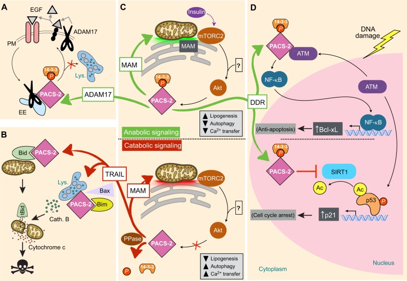
Fig. 4.
The PACS-2 Akt site and NLS together modulate membrane traffic, TRAIL-induced apoptosis, MAM integrity and the response to DNA damage. (A) Protein trafficking. Akt-phosphorylated pSer437-PACS-2 (pPACS-2) interacts with ADAM17 on early endosomes (EE) and mediates delivery of the protease to the cell surface where it sheds EGF ligands to stimulate EGFR signaling. In the absence of PACS-2, ADAM17 is degraded in lysosomes (Lys.). (B) TRAIL-induced MOMP. TRAIL triggers dephosphorylation of PACS-2 Ser437, which mediates two trafficking steps required for MOMP. In one trafficking step, PACS-2 binds full-length Bid and translocates Bid to mitochondria. In the other trafficking step, PACS-2 forms a complex with Bim and Bax on lysosomes called the PIXosome, which is required for lysosome membrane permeabilization to release cathepsin B (cath. B). (C) MAMS. Top panel: insulin or growth factors trigger activation of mTORC2 on mitochondria-associated membranes (MAMs; green shading at the ER–mitochondria contact site), which activates Akt to phosphorylate PACS-2. In turn, pPACS-2 increases MAM contacts, which may modulate ER–mitochondria exchange and support increased lipogenesis. The ? denotes signaling pathways that may lead to Akt-dependent phosphorylation of PACS-2 independent of MAM-localized TORC2. Bottom panel: in starved cells or cells treated with TRAIL, Akt is inhibited and PACS-2 Ser437 is dephosphorylated by a protein phosphatase (PPase). Dephosphorylated PACS-2 in turn remodels MAMs (red shading at the ER–mitochondria contact site), which may reduce lipogenesis but increase ER–mitochondrial Ca2+ exchange as well as induction of autophagy. (D) DNA damage response. Top panel: to support induction of the NF-κB and Bcl-xL anti-apoptotic pathway, cytoplasmic PACS-2 interacts with a pool of ATM released from the nucleus and maintains the DNA damage kinase in the cytoplasm. The cytoplasmic ATM then triggers activation of the canonical IκBα–NF-κB pathway that leads to induction of anti-apoptotic Bcl-xL. Bottom panel: to support induction of the p53–p21 cell cycle arrest pathway, pPACS-2 traffics to the nucleus where it binds and inhibits SIRT1 to protect acetylation of p53 bound to the p21 promoter, promoting p21 induction and cell cycle arrest. Green arrows, pro-survival-anabolic pathways mediated by pPACS-2. Red arrows, apoptotic or catabolic pathways mediated by dephosphorylated PACS-2. Ac, acetylation; DDR, DNA-damage response.

