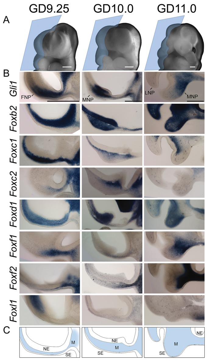
Fig. 3.

Fox gene expression during upper lip development. (A) Oblique images of untreated GD9.25, GD10 and GD11 embryos illustrate the plane of section (blue trapezoids) in the panels beneath. (B) Representative images are shown for n=3 embryos from at least two independent litters per gene per stage. Gli1 and seven Fox genes are expressed in the mesenchyme of the tissues that form the upper lip, i.e. the FNP at GD9.25 and MNP at GD10 and GD11. (C) Schematic showing tissue compartments of the developing face and brain at GD9.25, GD10 and GD11. FNP, frontonasal prominence; MNP, medial nasal process; SE, surface ectoderm; M, mesenchyme; NE, neuroectoderm. Scale bars: 200 μm.
