Fig. 7.
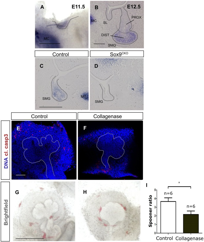
Type II collagen (Col2a1) is expressed in the distal progenitors and acts downstream of Sox9 possibly by contributing to branching. (A,B) In situ hybridisation for Col2a1 at the placode (A) and bud stage (B). (C,D) In situ hybridisation for Col2a1 at the bud stage (E12.5) in control (C) and Sox9CKO (D) submandibular glands. (E,F) Immunofluorescence for cleaved caspase 3 (red) in control (E) and collagenase-treated (F) submandibular gland explants. DNA is shown in blue (DAPI). (G,H) Brightfield images of control (G) and collagenase-treated (H) submandibular gland explants. (I) Spooner ratio of the number of buds produced in the control and collagenase-treated submandibular gland explants. *P<0.05. Dotted lines (A-H) delineate the salivary gland epithelium. Error bars represent s.e.m. DIST, distal; MC, Meckel's cartilage; PROX, proximal; SL, sublingual gland; SMG, submandibular gland. Scale bars: 250 μm (A); 50 μm (B); 100 μm (C-F); 500 μm (G,H).

