Fig. 2.
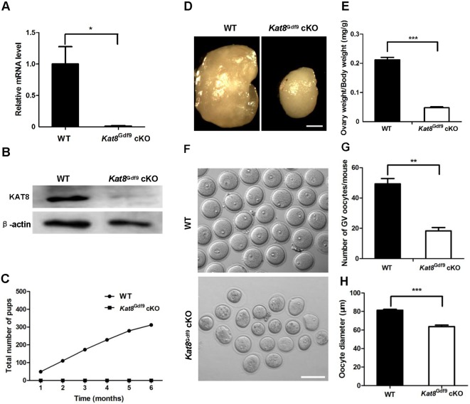
Kat8Gdf9 cKO mice display abnormal oogenesis and sterility. (A) Real-time PCR analysis of Kat8 mRNA levels in oocytes from 2-week-old WT and Kat8Gdf9 cKO mice. Kat8 mRNA levels in WT oocytes were arbitrarily set to 1, and values were normalized and plotted against Actb. (B) Western blot analysis of KAT8 in oocytes from 2-week-old mice. Per sample, ∼200 oocytes were used; β-actin served as a protein loading control. Images shown are representative of three independent experiments. (C) Fertility test. The cumulative number of pups obtained from matings of eight WT and eight Kat8Gdf9 cKO females to WT males over a 6 month period. (D) Representative images of ovaries from 8-week-old females. Scale bar: 0.5 mm. (E) Ratio of ovary weight to body weight in 8-week-old mice. (F) Representative images of GV stage oocytes from 8-week-old mice. Scale bar: 200 μm. (G) Mean number of GV stage oocytes obtained per mouse after priming with PMSG. (H) Mean diameter of GV stage oocytes. (A,E,G,H) Data are mean±s.e.m. from three independent experiments. *P<0.05, **P<0.01, ***P<0.001, t-test.

