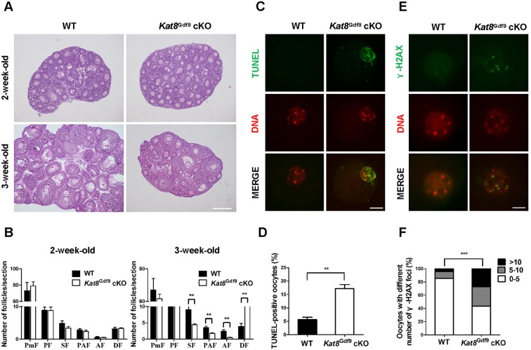
Fig. 3.
Kat8 deletion in oocytes causes follicle loss and oocyte defects. (A) Histological sections of ovaries from 2- and 3-week-old females stained with Hematoxylin and periodic acid-Schiff (PAS) reagent. (B) Quantitative analysis of follicles at different stages in ovary sections from 2- and 3-week-old WT and Kat8Gdf9 cKO females. PmF, primordial follicles; PF, primary follicles; SF, secondary follicles; PAF, preantral follicles; AF, antral follicles; DF, degenerating follicles. (C) Detection of apoptosis by TUNEL assay in oocytes from 3-week-old mice. (D) Percentage of TUNEL-positive oocytes. (B,D) Data are mean±s.e.m. from three independent experiments. **P<0.01, t-test. (E) Visualization of DSBs in oocyte nuclei from 3-week-old mice by γ-H2AX staining. (F) Proportions of WT and Kat8Gdf9 cKO oocytes with various numbers of γ-H2AX foci per nucleus. ***P<0.001, Chi-square test. Images (A,C,E) are representative of three independent experiments. Scale bars: 200 μm in A; 20 μm in C; 10 μm in E.

