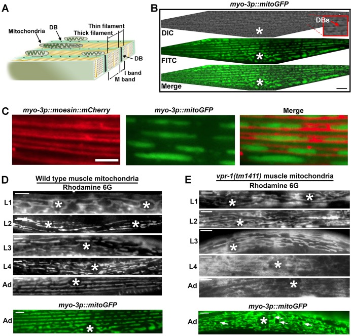
Fig. 1.
Mitochondrial organization in C. elegans body wall muscle. (A) Diagram of adult muscle myofilaments showing positions of mitochondria relative to I-bands. Mitochondrial tubules lie on top (or beneath, depending on orientation) of dense bodies (DBs). (B) Mitochondria in a single adult body wall muscle visualized with the myo-3p::mitoGFP transgene (Labrousse et al., 1999). Dense bodies are visible as dark dots along the muscle striations in the differential interference contrast (DIC) channel (arrow in higher magnification inset). FITC, fluorescein isothiocyanate. (C) Organization of mitochondria along thin filaments. The myo-3p::moesin::mCherry transgene labels muscle actin. Mitochondrial tubules extend along thin filaments, undergoing fission and fusion with adjacent tubules (Han et al., 2012). (D,E) Developmental timecourse of mitochondrial organization in wild-type (D) and vpr-1(tm1411) null mutant (E) muscles. Mitochondria are visualized by mitoGFP and Rhodamine 6G. Arrows indicate fat droplets; asterisk, nucleus; L1-L4, larval stages; Ad, adult stage. Scale bars: 5 µm.

