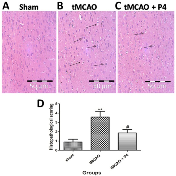
Fig. 7.

Effect of P4 on histopathology. (A-C) Representative histopathological photomicrograph of frontoparietal layers of the sham, tMCAO and tMCAO+P4 group. In the sham group, there was no vacuolation or any neuronal loss. In the tMCAO group, there was vacuolation and heavy neuronal loss. In the P4 treated group there was the partial neuronal loss. Magnification at 40×. (D) Histological alterations are represented graphically, showing significant changes (**P<0.001) in tMCAO and that, in the P4 treated group, there was a significant improvement (#P<0.05).
