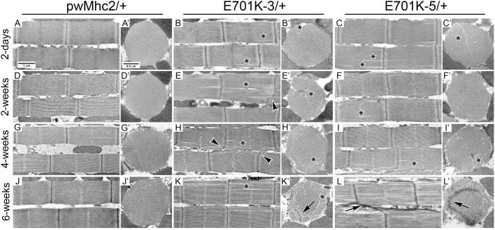
Fig. 2.
Ultrastructure of myofibrils from control and E701K/+ heterozygote IFMs during aging shows progressive defects in filament arrangement and Z-line organization. Transmission electron micrographs of longitudinal and transverse sections through IFM myofibrils from adult flies aged 2 days, 2 weeks, 4 weeks or 6 weeks after eclosion. PwMhc2 wild-type transgenic controls (A,A′,D,D′,G,G′,J,J′) assemble into well-organized sarcomeres (A) and this structure is retained as the flies age (D,G,J). PwMhc2 myofilaments are packed in a rigid double hexagonal array (A′) that is consistent during aging (D′,G′,J′). Two-day-old myofibrils from E701K-3/+ and E701K-5/+ IFMs also assemble well-ordered sarcomeres (B,C) with double-hexagonal filament packing (B′,C′), although occasional minor gaps in the microfilament arrays are observed (asterisks). These gaps are exacerbated during aging in longitudinal sections (E,F,H,I,K,L) and are particularly evident in transverse sections (E′,F′,H′,I′,K′,L′). As the heterozygotes age, Z-lines become non-linear (arrowheads), and Z-line streaming, where Z-line material is mislocalized, is observed (K′,L,L′; arrows). Scale bars: 1 µm (longitudinal sections); 0.5 µm (transverse sections).

