Fig. 4.
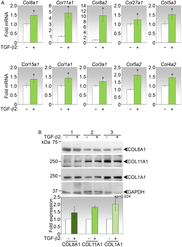
Induction of top collagen genes by TGFβ2 in primary mouse conjunctival fibroblasts. (A) Real-time PCR analyses of collagen genes in fibroblasts stimulated for 72 h with TGFβ2. Data shown are representative of three independent experiments, and show mean fold-change±s.d. relative to untreated controls. *P<0.05 for fold-change in treated versus control. (B) Immunoblot analyses of COL8A1, COL11A1 and COL1A1 in mouse fibroblasts treated as indicated for 72 h. Three independent sets of experiments are shown. Fold-change in expression in TGFβ2-treated relative to control cells, both normalized to GAPDH, and P value (corrected by Bonferroni adjustment) where significant, are shown in the densitometric analyses below the immunoblot.

