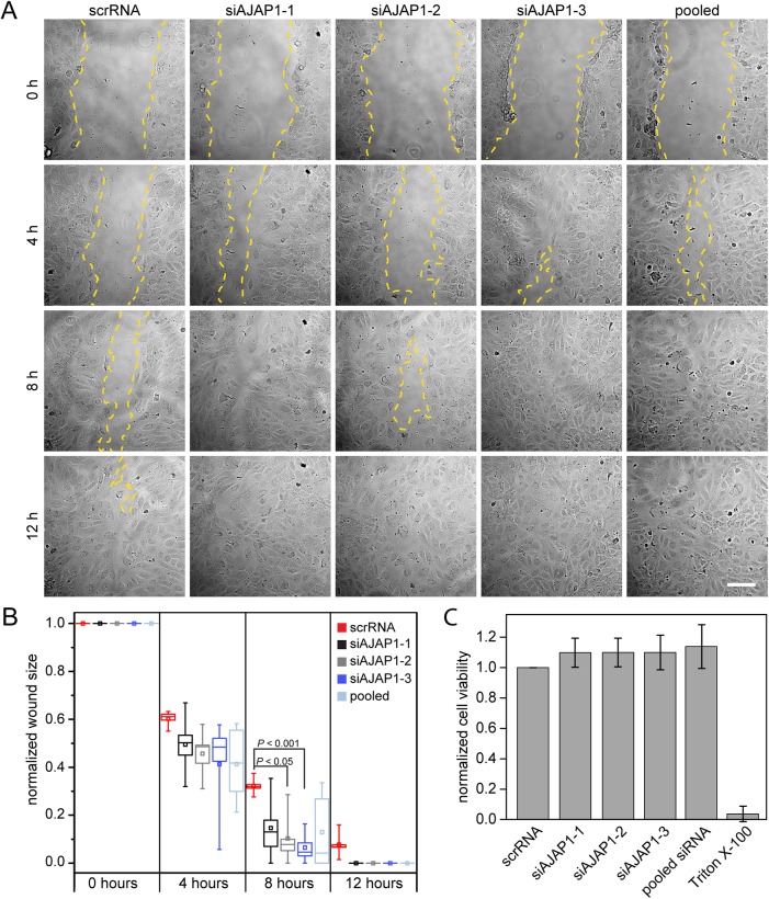
Fig. 2.
AJAP1 knockdown induces wound closure in HUVECs. (A) Migration of scrRNA, siAJAP1-1, siAJAP1-2, siAJAP1-3 and pooled siRNA transfected HUVECs in an in vitro wound healing assay at different time points after wounding. The yellow dashed line indicates the migrating front. Microscope: Zeiss Axio Observer.Z1; objective lens: Fluar 10×/0.5; scale bar: 100 µm. (B) The normalized wound size is plotted for each time point and each condition. The wound size decreases faster over time when AJAP1 is down regulated. Five independent experiments were performed per condition. Data is normalized to the scratch size at time point 0 for each condition. Only samples with a comparable wound area size were chosen for the analysis. The box contains 50% of the data points, the middle line of the box is the median and the square is the arithmetic mean. Whiskers represent minimum/maximum values. Statistics were performed using the t-test with the Bonferroni correction. (C) HUVECs were transfected with scrRNA or siAJAP1. Cell viability was measured by a MTS assay and is expressed as a percentage of scrRNA transfected cells. Mean±s.d. n=3.

