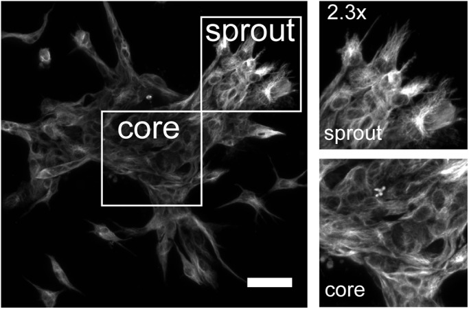
Fig. 4.

AJAP1 remains localized in fibrillar structures during HUVEC spheroid sprouting. The maximum Z-projection shows an immunofluorescence staining of a sprouting spheroid with the localization of AJAP1 (antibody: Abcam). Focusing on the core and the sprouting region, AJAP1 always appears in fibrillar structures indicating that its localization does not alter in polarizing and migrating cells. Microscope: LSM 780; objective lens: LD EC Epiplan-Neofluar 20×/0.22, spacing: 1.58 µm; scale bar: 25 µm.
