Fig. 1.
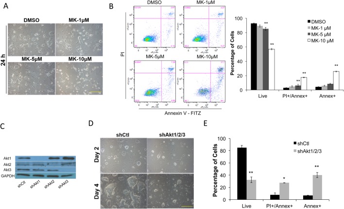
Blocking Akt activity leads to ESC apoptosis. (A) R1-ESCs grown in 2i/LIF medium plus DMSO control or 1–10 µM of MK for 24 h. Scale bar: 250 µm. (B) R1 ESCs treated as described in A were incubated with Annexin V-FITC (Annex) and Propidium Iodide (PI) for 30 min and analyzed by flow cytometry. Percentages of late apoptotic/necrotic (Annex+/PI+), early apoptotic (Annex+), and live cells (unstained) are shown (data shown are mean±s.d., **P<0.01, n=3). (C) Western blot of total protein extracts from ESCs 3 days after lentiviral control (shCtl), shAkt1, shAkt2, and shAkt3 transduction, respectively. Proteins were blotted with the respective Akt antibodies with GAPDH used as a loading control. (D) ESCs expressing lentiviral shCtl or a combination of shAkt1, shAkt2, and shAkt3 (shAkt1/2/3) for 2 and 4 days, respectively, in 2i/LIF medium. Scale bar: 250 µm. (E) ESCs treated as described in D were incubated with Annex and PI for 30 min and analyzed by flow cytometry. Percentage of early, late apoptotic and live cells were determined as described in B (data shown are mean±s.d., *P<0.05, **P<0.01, n=2).

